This web page was produced as an assignment for Genetics 564, an undergraduate course at the University of Wisconsin-Madison.
Disease Overview:
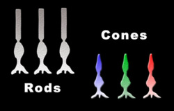
Achromatopsia is a vision disorder that causes various phenotypic problems in the retina. [1] The retina contains both cones and rods that are essential to correct vision. Rods help with the ability to see in dim or dark lighting, but they do not play a role in providing color details or sharp vision. The cones located in the retina are responsible for seeing color and details. There are three different types of cones. The protan class is sensitive to long wave lengths (red spectrum), the deutan cones are sensative to middle length wavelengths (green spectrum), and the tritan cones are sensitive to short wave lengths (the blue spectrum). These cones work simultaneously and contribute to one another allowing the eye to see a very wide range of colors. [4]
Achromatopsia is caused by the absence of these colored cones in the retina. [4] As a result of the non-functioning cones, in all cases Achromatopsia causes a form of colorblindness. In addition to the deficiency in the ability to distinguish certain colors, this disease also causes other symptoms that can be related to the loss of cone function; for example: decreased vision accuracy (cones contribute to sharp vision) and light sensitivity. [1] This disease is also associated with nystagmus (shaking in the eyes) and lazy eyes. [2] Achromatopsia is not the same as red-green color blindness and is also much less common than red-green colorblindness. [2]
|
The most common form of Achromatopsia is
Complete Achromatopsia - loss of function in all three cones, however
there are rare cases of Incomplete Achromatopsia where only one or two
of the cones in non-functioning. [2]
Achromatopsia occurs in approximately one in 40,00 live births. Its prevalence varies throughout the world because of the diseases genetic nature. It is most common in regions in the world that have high rates of marriages between relatives. [2] Achromatopsia is an autosomal recessive disease. [4] With this type of inheritance pattern the child receives one copy of the gene from each parent. If neither parent is affected nor a carrier, the child will not have the disease. If both parents have the disease then all of their offspring will also be affected. As a result of the rarity of this disease it is often difficult for patients to receive the correct diagnosis and adequate treatment. |
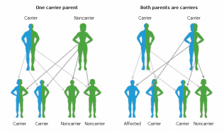
Figure 2: Examples of autosomal recessive inheritance
|
Diagnosis:
There are many factors that are considered when diagnosing this disease. These factors include the patients medical history, color vision testing,
electrophysiologic examination, and examination of family history (often with a genetic counselor). [4]
There are many factors that are considered when diagnosing this disease. These factors include the patients medical history, color vision testing,
electrophysiologic examination, and examination of family history (often with a genetic counselor). [4]
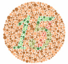
Color Vision Testing:
The overarching phenotype of Achromatopsia is the lack of reliable color perception. In most cases, people with Achromatopsia have a deficiency in all in all three axes of color vision that correspond to thre three classes of cones that are discussed above. The most important type of color vision test and the test that yields that most information that is of use for diagnosis is the Red- Green color test that is done. [4]
The overarching phenotype of Achromatopsia is the lack of reliable color perception. In most cases, people with Achromatopsia have a deficiency in all in all three axes of color vision that correspond to thre three classes of cones that are discussed above. The most important type of color vision test and the test that yields that most information that is of use for diagnosis is the Red- Green color test that is done. [4]
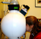
Electrophysiological Examination (ERG):
This is a noninvasive test that consists of a either single bright flash or a series of light "flickers". [4] The exam shows the retinas electrical response to a light stimulus, evaluating the function of the retina. Surpressing the eyes rod photoreceptors with the bright background allows the cone photoreceptor response to be isolated. [6] In the single flash ERG people with Achromatopsia typically have an absent or diminished photopic response. When a flicker ERG is conducted typically people with Achromatopsia do not respond with the typical cone drive gast pathway response that would normally be elicited by high flash intensities. [6]
This is a noninvasive test that consists of a either single bright flash or a series of light "flickers". [4] The exam shows the retinas electrical response to a light stimulus, evaluating the function of the retina. Surpressing the eyes rod photoreceptors with the bright background allows the cone photoreceptor response to be isolated. [6] In the single flash ERG people with Achromatopsia typically have an absent or diminished photopic response. When a flicker ERG is conducted typically people with Achromatopsia do not respond with the typical cone drive gast pathway response that would normally be elicited by high flash intensities. [6]
Treatment:
There is currently no cure or medical treatment for Achromatopsia, but there are various techniques for managing the disease. THese techniques are also very important for preventing any further damage to the retina from occurring. [4]
There is currently no cure or medical treatment for Achromatopsia, but there are various techniques for managing the disease. THese techniques are also very important for preventing any further damage to the retina from occurring. [4]
- Surveillance of the disease from a physician is very important. Children with the disease should have an ophthalmologic exam every 6-12 months in-order to monitor changes in refraction and allow the child's eye to achieve its maximum visual acuity possible. Adults should be examined every two to three years. [4]
- There are dark tinted glasses and red tinted contacts that can be worn to help prevent sensitivity to bright light. [4] These glasses/contacts also can improve visual accuracy. [7]
- Avoiding circumstances that are dangerous to the eyes retina is also encourage. Avoiding extreme bright lights and ensuring the use of dark tinted glasses when in bright lights is essential to avoiding further damage and being comfortable. [7]
- There are other possibilities of management such as tinted computer screens [3], sitting in the front of the class room [7], etc.
CNGA3 Gene & Protein and Achromatopsia:
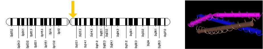
Figure 3: The image of the left shows where the gene is located on the 2nd chromsome. The image on the right demonstrates the 3-D structure of the CNGA3 protein
Cyclic-nucleotide-gated channel alpha 3 (CNGA3) is one gene that studies have linked to the inheritance of Achromatopsia. CNGA3 is a gene that is responsible for encoding proteins and more specifically encodes a family of proteins that are required for normal and olfactory signal transduction. [8] CNGA3 is located near the centromere on the second chromosome.[5] Over 50 different mutations located on this gene have been linked to color deficiency problems and most commonly linked to Complete Achromatopsia. The majority of these mutations change only one amino acid in the CNGA3 protein. This change alters the structure of CNG channels associated with this protein so that it can no longer regulate the flow of ions into the cones in the retina. Normal cone function is essential for color vision, sharpness so these mutations affect how people see. [9]
Resources:
[1] http://omim.org/entry/216900
[2] http://www.aapos.org/terms/conditions/10
[3] http://optometry.berkeley.edu/focus_magazine/focus02_achromat.html
[4] http://www.ncbi.nlm.nih.gov/books/NBK1418/
[5] Wissinger, B., Gamer, D., Jagle, H., Giorda, R., Marx, T., Mayer, S., Tippmann, S., Broghammer, M., Jurklies, B., Rosenberg, T., Jacobson, S. G., Sener, E. C., and 17 others. CNGA3 mutations in hereditary cone photoreceptor disorders. Am. J. Hum. Genet. 69: 722-737, 2001. [PubMed: 11536077, related citations] [Full Text: Elsevier Science]
[6] Fatih Cakir Gundogan, Ahmet Tas and Gungor Sobaci (2011). Electroretinogram in Hereditary Retinal Disorders, Electroretinograms, Dr. Gregor Belusic (Ed.), ISBN: 978-953-307-383-5, InTech, Available from: http://www.intechopen.com/books/electroretinograms/electroretinogram-in-hereditary-retinal-disorders
[7] Park WL, Sunness JS. Red contact lenses for alleviation of photophobia in patients with cone disorders. Am J Ophthalmol. 2004;137:774–5. [PubMed]
[8] http://www.ncbi.nlm.nih.gov/gene?cmd=Retrieve&dopt=full_report&list_uids=1261
[9] http://ghr.nlm.nih.gov/gene/CNGA3
|
|
Created by: Alison Heydorn University of Wisconsin Last Date Modified: 05/16/2014 |
